Cysts are closed capsule or sac-like structures, typically filled with liquid, semisolid, or gaseous material, very much like a blister. In this article, we will describe the various types.
This occurs within the tissue and can affect any part of the body. They vary in size from microscopic to the size of some team-sport balls – large blisters can displace internal organs.
In anatomy, a cyst can also refer to any normal bag or sac in the body, such as the bladder. This article refers to an abnormal sac or pocket in the body that contains liquid, gaseous, or semisolid substances.
A cyst is not a normal part of the tissue where it is located. It has a distinct membrane and is separated from nearby tissue – the outer (capsular) portion of a cyst is called the cyst wall. If the sac is filled with pus it is not a cyst; it is an abscess.
What causes cysts?
Common causes include:
- tumours
- genetic conditions
- infections
- a fault in an organ of a developing embryo
- a defect in the cells
- chronic inflammatory conditions
- blockages of ducts in the body that cause fluids to build up
- a parasite
- an injury that breaks a vessel
Benign and malignant cysts
Most blisters are benign and are caused by blockages in the body’s natural drainage systems. However, some may be tumours that form inside tumours – these can potentially be malignant. Examples include keratocysts and dermoid cysts.
Signs and Symptoms of cysts
Signs and symptoms vary enormously depending on what type of cyst it is. In many cases, a person becomes aware of an abnormal lump, particularly in cases with blisters of the skin or just below the skin. A person may notice it in their breasts when they examine them by touching them. Breast cysts are often painful.
Some blisters in the brain can cause headaches, as well as other symptoms.
Many internal cysts, such as those in the kidneys or the liver, may not have any symptoms and go unnoticed until an imaging scan (MRI scan, CAT scan, or ultrasound) detects them.
Types of cysts
Some of the most common types are listed below:
- Acne Cysts
Cystic, or nodulocystic, acne is a severe type of acne in which the pores in the skin become blocked, leading to infection and inflammation.
- Arachnoid Blisters
The arachnoid membrane covers the brain. During fetal development, the arachnoid membrane doubles up or splits to form an abnormal pocket of cerebrospinal fluid. In some cases, doctors need to drain it out. It may affect newborn babies.
- Baker’s blisters
It is also called popliteal cysts. A person with this type of disease often experiences a bulge and a feeling of tightness behind the knee. Pain gets worse when extending the knee or during physical activity. It can cause knee joints, such as arthritis or a cartilage tear.
- Bartholin’s cysts
These may occur if the ducts of the Bartholin glands (situated inside the vagina) become blocked. Women may undergo surgery and/or be prescribed antibiotics.
- Chalazion blisters
Very small eyelid glands (meibomian glands) make a lubricant that comes out of tiny openings in the edges of the eyelids. It can form if the ducts are blocked.
- Colloid blisters
These are blisters that contain gelatinous material in the brain. In most cases, the recommended treatment is surgical removal.
- Dentigerous cysts
It surrounding the crown of an unerupted tooth.
- Dermoid cysts
This type includes mature skin, hair follicles, sweat glands, clumps of long hair, as well as fat, bone, cartilage, and thyroid tissue.
- Epididymal cysts
These are blisters (spermatocele) that form in the vessels attached to the testes. This type of disease is estimated to affect 20-40 per cent of American males and does not typically impair fertility or require treatment. If it causes discomfort a doctor may suggest surgery.
- Hydatid Blisters
A relatively small tapeworm forms sacs in the lungs or liver. Treatment includes surgery and medication.
- Pancreatic Cysts
They are referred to as pseudocysts as they do not contain the type of cells found in true cysts. They can include cells normally found in other organs, such as the stomach or intestines.
- Periapical cysts
These are also known as radicular cysts. They are the most common odontogenic (relating to the formation and development of teeth) blister and are usually caused by inflammation of the pulp, pulp death, or dental caries.
- Pilar Cysts
These are also known as trichilemmal cysts. They are fluid-filled that form from a hair follicle and are most commonly found in the scalp.
- Pilonidal Blisters
This disease form in the skin near the tailbone (lower back), and can sometimes contain ingrown hair. This type can grow in clusters, which sometimes create a hole or cavity in the skin.
- Renal cysts (kidneys)
Several types can develop in the kidneys. Solitary blisters contain fluids and may sometimes include blood. Some are present at birth; others may be caused by tubular blockages. People with kidney vascular diseases may have cysts formed by the dilatation of blood vessels.
- Pineal gland cysts
These are benign cysts that form in the pineal gland in the brain. According to autopsy records, pineal gland blisters are fairly common.
- Sebaceous Cysts
The skin is lubricated by sebaceous fluid, which can build up inside a pore or hair follicle and form a lump filled with thick, greasy substances. Sebaceous blisters are most commonly found on the skin of the face, back, scalp, and scrotum.
- Tarlov cysts
These are also known as perineural/perineurial cysts, as well as sacral nerve root cysts. It is located at the base of the spine and is filled with cerebrospinal fluid.
- Vocal fold cysts
There are two types – mucus retention and epidermoid cysts. Vocal fold cysts can interfere with the quality of the person’s speech, sometimes causing vocal cords to produce multiple tones simultaneously (diplophonia), or hoarseness and breathy speech (dysphonia).
Treatments for cysts
The Treatment will depend on various factors, including the type of the disease, where it is, its size, and the degree of discomfort it is causing.
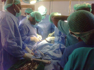
A very large cyst that causes symptoms can be surgically removed. Sometimes, the doctors may decide to drain or aspirate it by inserting a needle or catheter into the cavity.
If it is not easily accessible, drainage or aspiration is often done with the help of radiologic imaging so that the doctor can accurately guide the needle/catheter into the target area.
Sometimes, doctors examine the aspirated liquid under a microscope to determine whether cancerous cells are present.
If doctors suspect that your disease might be cancerous, it may be removed surgically, or they may order a biopsy of the capsule (cyst wall).
Many cysts arise as a result of a chronic or underlying medical condition, as may be the case with fibrocystic breast disease or polycystic.
References
Retrieved from bestpractice Pu, Y., Mahankali, S., Hou, J., Li, J., Lancaster, J. L., Gao, J. H., … Fox, P. T. (2007, September 20).
High prevalence of pineal cysts in healthy adults demonstrated by high-resolution, noncontrast brain MR imaging. American Journal of Neuroradiology, 28(9), 1706-1709. Retrieved from Woodward, P. J., Schwab, C. M., & Sesterhenn, I. A. (2003, January). From the archives of the AFIP: Extratesticular scrotal masses: radiologic-pathologic correlation.


